


Healthy vision

Vision disorders

Vitreous opacities
Vitreous opacities are caused by small irregular bodies which float in the vitreous body of the eye. They are called floaters.
What’s the problem?
VITREOUS BODY
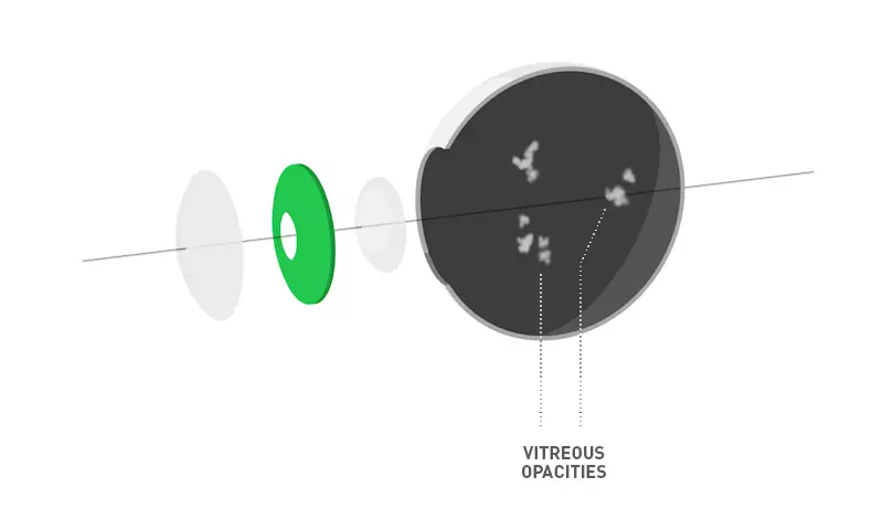
The vitreous body is a jelly-like filling of the eyeball, reminiscent of a raw egg white, which helps it keep its shape. Vitreous opacities most frequently occur when looking at a white surface or at the light. When the vitreous opacities move, they appear in the field of vision.
They can have the shape of specks, stains, threads or cobwebs.
They increase mainly with age, but we can also encounter them in younger patients also after accidents or in the case of short-sightedness. In most cases they are harmless, but they can have a very unpleasant impact on the patient’s life.
What happens during surgery?
LASER VITREOLYSIS
Laser vitreolysis is a non-invasive, painless procedure which is carried out using a special YAG laser and helps remove smaller, localised vitreous opacities.
The eye is first desensitised using drops, then a stabilising contact lens is applied. This keeps the eye open and reduces the speed of its movements. The patient follows the doctor’s instructions and looks at various points in order to remove all localised opacities.
During the procedure, opacities are surveyed using a focusing ray and subsequently removed.
Once the laser ray has been aimed, a micro-explosion of plasma is caused in the vitreous body, which transforms everything nearby into gas. The obstructing shapes disappear or are broken up into smaller pieces and moved to the edge of the field of vision where they cannot be seen.
The procedure takes 15 to 60 minutes and can be performed on both eyes within one day.
The laser treatment usually needs to be repeated on the eye several times, and thus the treatment may include several sessions.
- Laser vitreolysis is not suitable for patients with very dense and large vitreous opacities, infection-related vitreous opacities and opacities located in unsuitable positions (very close to the retina or lens)
- The procedure is usually performed in the central sections of the vitreous body.
- The procedure is performed using a local anaesthetic and is painless.
What can I expect after the procedure
and what post-surgery care is required?
Immediately following the procedure, blurred vision can occur for 3 to 4 hours due to the widening of the pupil and the use of the contact lens
In rare cases, the patient may have the feeling of the presence of a foreign body in the eye, and the eye may run. This feeling results from the slight irritation to the eye caused by the contact lens used during the procedure. These problems usually last only a few hours.
During the post-surgery period, treatment with drops is not usually needed, and no restrictions are required.

Equipment and instruments
-
Surgical microscope ZEISS Lumera
Surgical microscope ZEISS Lumera
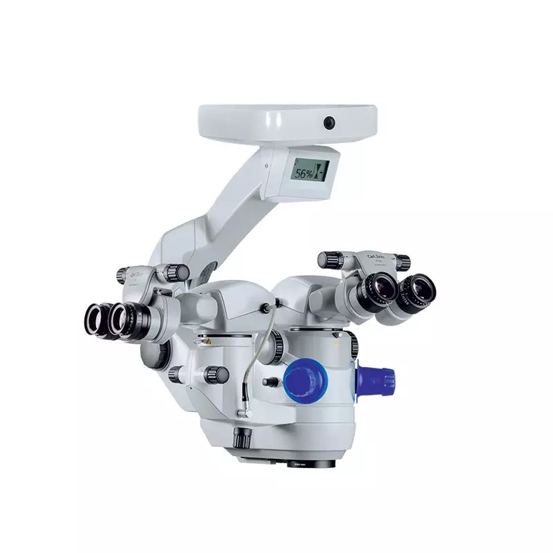
At present the best surgical microscope for eye micro-surgery in the world. In the operating theatre, a unique 3D HD stereoscopic system is also installed with the microscope, enabling transmission of the operations, their recording and imaging in 3D HD (three-dimensionally). This system is unique in Europe and was specially developed for the needs of the UVEA Mediklinik eye clinic.
-
Callisto Eye navigation system
Callisto Eye navigation system
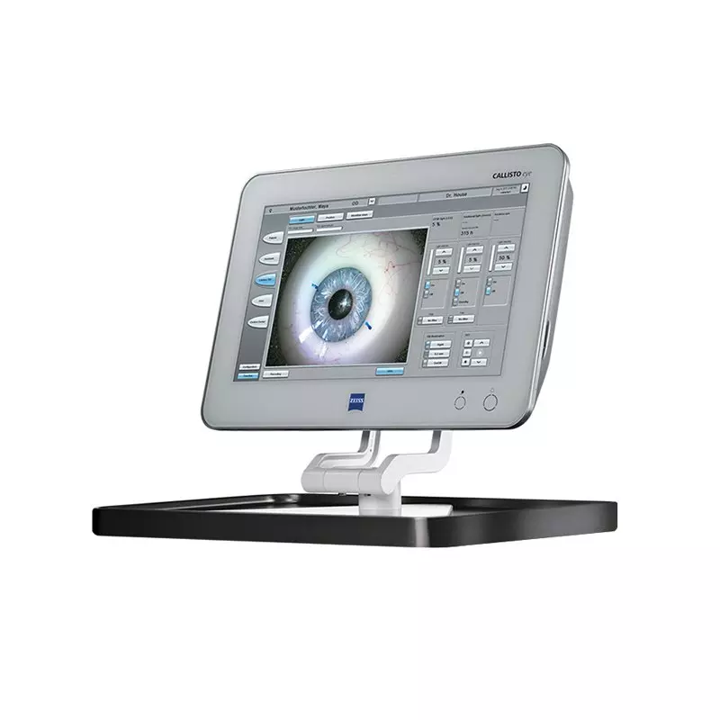
Callisto Eye is a unique eye monitoring system. It was the first to be installed in the Czech Republic and Slovakia, and right in the UVEA Mediklinik eye clinic in Martin. For patients, it is an irreplaceable guarantee of safety and precision, thanks to its live transfer of localisation marks into the surgeon’s microscope during intraocular operations, such as cataract operations or ceratoplastic surgery. The Callisto eye navigation system is capable of monitoring and adapting to eye movements in real time during the operation.
-
Constellation phacoemulsification device
Constellation phacoemulsification device
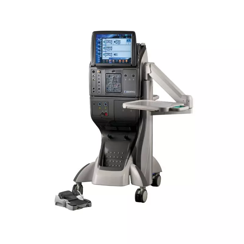
This is a multifunctional surgical device which enables us to perform operations on the anterior and posterior eye segment. We use it for the emulsification of the intraocular lens or for operations of the vitreous body and retina, even in the most complicated cases. The emulsification of an opaque or clear intraocular lens takes place with the use of a very low ultrasound energy, thus leading to a minimal traumatisation to eye structures during the operation.
The device is equipped with
A torso mode of phacoemulsification
Integrated xenon light source
High-speed vitrectomy system
Integrated endolaser
-
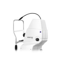
Funduskamera Visucam 500
Funduskamera Visucam 500
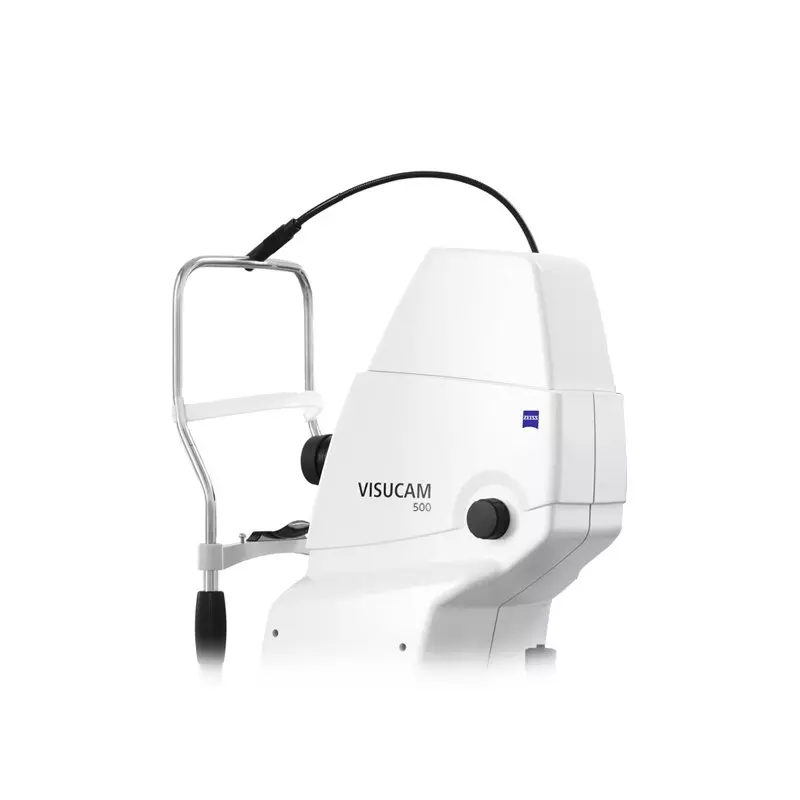
The camera VISUCAM 500 with its 24-megapixel sensor creates brilliant, detail-rich images which are an effective help in the diagnostics and monitoring of a wide range of eye diseases: from glaucoma and diabetic retinopathy through to age-related macular degeneration (ARMD).
The advantage of the Visucam camera is the high definition imaging of the retina and its wide range of imaging regimes -
Cirrus HD coherent tomograph
Cirrus HD coherent tomograph
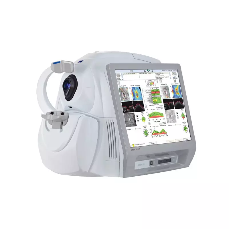
At the UVEA Mediklinik, we have the Cirrus coherent tomograph which is one of the most modern, highly useful examination and imaging device aimed at the detailed examination of posterior and anterior eye structures such as the retina, optic nerve and cornea. We use it for rapid and simple diagnostics before most eye procedures. Thanks to its perfect optics, the Cirrus HD-OCT also offers our doctors highly professional analyses and graphical imaging of the results obtained. As a result, doctors can monitor the development of a wide range of eye diseases.
-
Cryomatic MK II
Cryomatic MK II
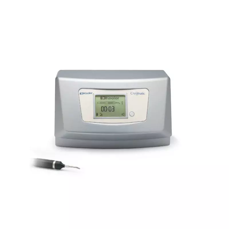
This is a new generation of cryosurgical device by Keeler, which stands out for its high reliability. This device is mainly used for operations of the posterior eye segment such as retinal tears. The device is equipped with a microprocessor which electronically controls the freezing and defrosting cycle.
By freezing the affected spot, we can ensure that the affected part of the retina remains firmly fixed. The device works with CO2 or N2O freezing medium.
Is surgery right for you?
To help you find your way around, we have prepared a brief overview of surgeries available for different types of refractory disorders or diseases. Find out which type of surgery is best for you.


Price list
To help you find your way around, we have prepared a brief overview of surgeries available for different types of refractory disorders or diseases. Find out which type of surgery is best for you.
